Get Back
to Your Life!
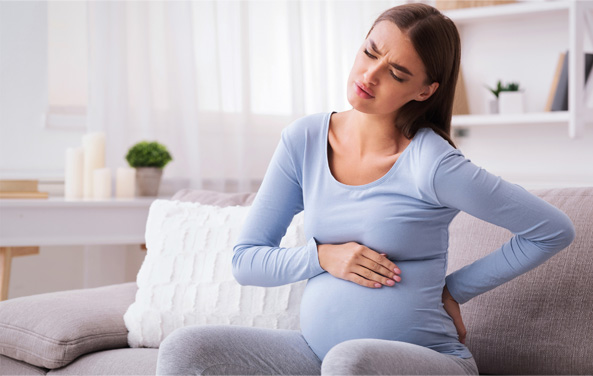
Are you experiencing low-back pain?
It could be sacroiliac (SI) joint dysfunction.
90%
of the adult population will experience lower back pain at some point in their lives.
25%
of these individual’s pain will be caused by sacroiliac (SI) joint dysfunction.2
This may be the type of pain you are experiencing. In most cases, there are certain health factors that may have contributed to this condition. The information provided here will help you learn more about SI joint dysfunction and present options that could help you get back to your life.
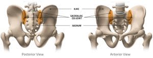
What is the SI joint and its primary function?
The sacroiliac joint or SI joint is the connection point between the sacrum (triangular bone at the bottom of the spine) and the pelvis (iliac bone or hip bone). Unlike most joints that slide and glide, the SI joint is considered somewhat static, although it does allow for a few millimeters of motion. This joint is often referred to as the body’s shock absorber, providing stability and load-bearing characteristics by transferring body weight and downward forces through the pelvis to the legs while engaging in physical activities like walking, running, climbing, jumping, etc. This unique functionality can eventually lead to the SI joints becoming strained, injured, or affected by a chronic condition. It can also become unaligned, exposing or pinching nerves and generating pain in your lower back and extremities.

Pain standing or sitting for long periods
Common SI Joint
Pain Indicators
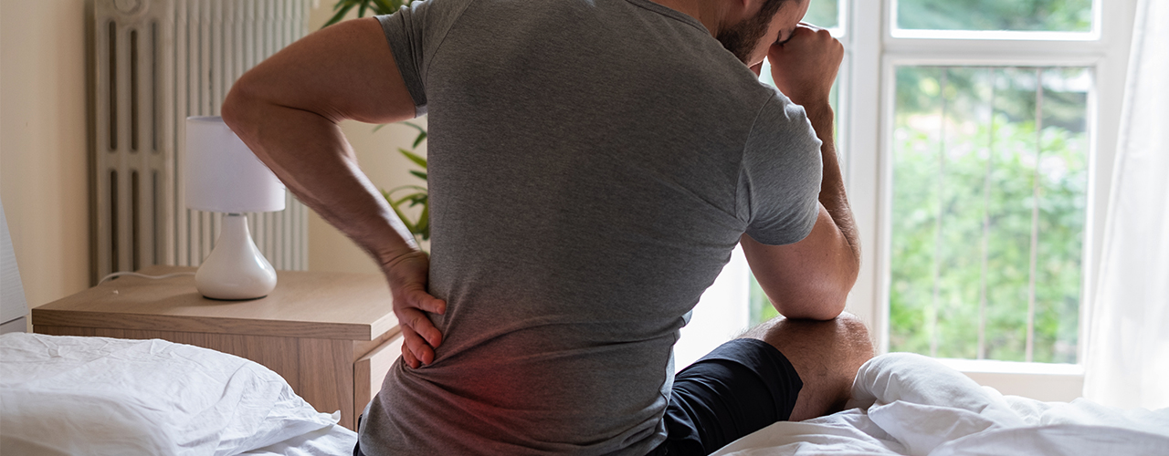
Pain and difficulty sleeping on one side or both

Pain while exercising or stretching
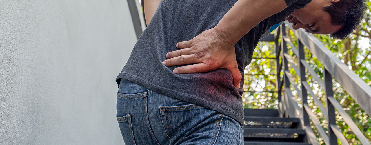
Pain and difficulty walking up or down stairs
Symptoms and Pain locations related to SI joint dysfunction:
- Lower back pain
- Sensation of pain, numbness, tingling, weakness in lower extremities
- Pelvic/buttock pain
- Hip/groin pain
- Leg instability (buckling, giving way)
- Disturbed sleep patterns due to pain
- Pain from sitting and/or standing from the seated position
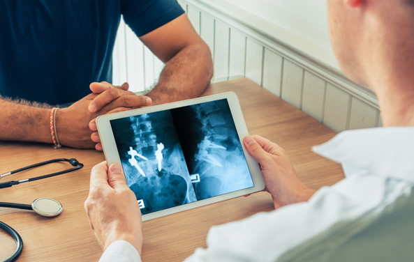
Pain in the lower back can originate from several pain generators, so it is essential to determine the source of your pain. Your physician may accomplish this through physical examination, radiographs, MRIs, and analgesic injections. Time spent with your physician reviewing the location of your pain and how you experience your pain is crucial.

What caused my SI Joint Pain?
There can be many causes of SI joint dysfunction, including but not limited to:


Like most other joints in the body, the SI joints have a cartilage layer covering the bone. This cartilage allows for some movement and acts as a shock absorber between the sacrum and iliac bones. When this cartilage becomes damaged or is worn away by age, the bones begin to rub on each other, and degenerative arthritis (osteoarthritis) occurs.
DEGENERATIVE ARTHRITIS

CLOSE X
During pregnancy your hip and sacroiliac joints will begin to naturally loosen. This is your body preparing to give birth. Add to that a change in the way some women walk as a result of pregnancy and that can cause your sacroiliac joints to become inflamed. This kind of pain can last through delivery and into the postpartum period.
PREGNANCY & NATURAL CHILD BIRTH
CLOSE X
Trauma caused by a series of small, repetitive injuries that accumulate over time, such as in the case of an athlete, or it could be the result of a sudden impact such as a pelvic fracture from a motor vehicle accident or fall.
TRAUMA

CLOSE X
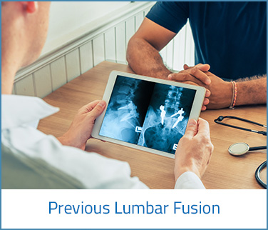
Fusion of the lumbar spine alters its natural movement and ability to absorb shock, resulting in increased load or stress on the adjacent lumbar segment and potential for degeneration. Sometimes this “transfers” forces to the SI joints, especially when prior lumbar fusion included the L5/S1 level.
PREVIOUS LUMBAR FUSION
CLOSE X
“Diagnosing SI joint dysfunction can be challenging. The lumbar spine, discs, sacrum, ilium and SI joints (your pelvis/hips) all share the transfer of load and stress to and from the legs, so it could be a combination of pain generators. Identifying the exact pain source is why multiple diagnostic tools are used by physicians to determine the cause of your pain.*”
DIAGNOSTIC TOOLS


Options for Treating Sacroiliac (SI) Joint Pain
- Medications
- Physical Therapy
- Therapeutic Injections
- Surgical Intervention
Only you and your physician know what is right for you and your specific diagnosis.
The information provided here is not intended to replace professional medical care.
Patient Testimonial: Charlotte’s Story
Disclaimer:
The material in this section of the website is intended for educational resource purposes only and is not meant to replace conversations between a patient and their physician or member of their health care team. Please consult with your physician to obtain a complete list of indications, contraindications, precautions, warnings, clinical results and other important medical information that pertains to this procedure. The decision to receive medical treatment is individualized to the patient and the patient’s symptoms. The information presented on this site may not apply to your condition, treatment or its outcome, as surgical techniques vary and complications can occur. It is important to discuss the viability of any surgical procedure with your physician to decide the right treatment option.
2. Bernard TN, Jr, Kirkaldy-Willis WH. Recognizing specific characteristics of nonspecific low back pain. Clin Orthop Relat Res. 1987;217:266–80.
We can help you find a doctor if you don’t have one and you think SI fusion might be right for you.


